
Skeda:DTI-sagittal-fibers.jpg
Madhësia e këtij shikimi: 643 × 600 pixel. Rezolucione të tjera: 257 × 240 pixel | 515 × 480 pixel | 1.021 × 952 pixel.
Dokument origjinal (1.021 × 952 pixela, madhësia e skedës: 294 KB, tipi MIME: image/jpeg)
|
Përmbledhje
| Përshkrimi |
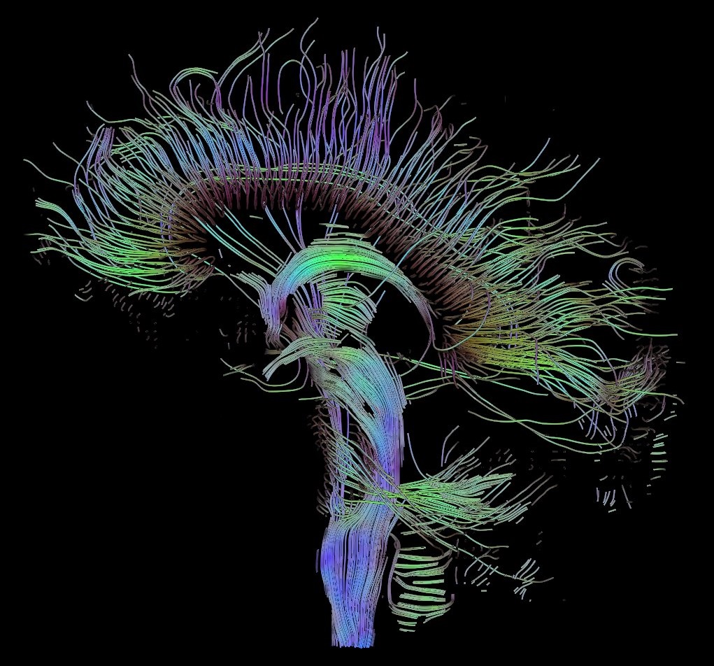
English: Visualization of a DTI measurement of a human brain. Depicted are reconstructed fiber tracts that run through the mid-sagittal plane. Especially prominent are the U-shaped fibers that connect the two hemispheres through the corpus callosum (the fibers come out of the image plane and consequently bend towards the top) and the fiber tracts that descend toward the spine (blue, within the image plane)
Français : Visualisation d'une mesure DTI d'un cerveau humain. Ce qui est représenté sont des faisceaux de fibres reconstruits qui traversent le plan demi-sagittal. On observe les fibres en U qui connectent les deux hémisphères à travers le corps calleux, qui sont particulièrement importantes (les fibres sortent du plan de l'image et par conséquent se courber vers le haut) ainsi que les faisceaux de fibres qui descendent vers la colonne vertébrale (bleu, dans le plan de l'image)
Deutsch: Traktographie-Verfahren rekonstruieren aus den Messdaten der Diffusions-Tensor-Bildgebung den anzunehmenden Verlauf größerer Nervenbahnen. Hier dargestellt sind die Ergebnisse für ein menschliches Gehirn; um die Übersichtlichkeit zu wahren, beschränkt sich die Abbildung auf Bahnen, die die Medianebene schneiden. Insbesondere sind dies die U-förmigen Faserbündel, die die beiden Hirnhälften verbinden (sie durchstoßen die Bildebene und sind nach oben gebogen) sowie die Faserbündel, die zum Rückenmark ziehen (blau dargestellt, liegen innerhalb der Bildebene) |
| Data | |
| Burimi | Punë e juaja |
| Autori | Thomas Schultz |
| Leja (Ripërdor këtë skedë) |
Rendering is own work, using a modified version of the BioTensor application developed at the University of Utah. The dataset is courtesy of Gordon Kindlmann at the Scientific Computing and Imaging Institute, University of Utah, and Andrew Alexander, W.M. Keck Laboratory for Functional Brain Imaging and Behaviour, University of Wisconsin, Madison. It is publicly available from [1] |
Licencim
Unë, krijuesi i kësaj pune, e publikoj këtu në bazë të licensës në vijim:

|
Ju jepet leje për ta kopjuar, shpërndarë dhe/ose ndryshuar këtë dokument sipas kushteve të Licencës GNU për Dokumentim të Lirë, versioni 1.2 ose çfarëdo versioni të mëpasshëm të botuar nga Free Software Foundation; pa Seksione të Pandryshueshme, pa Tekste Kapakësh të Përparmë, dhe pa Tekste Kapakësh të Pasmë. Një kopje e kësaj licence është përfshirë në seksionin e titulluar GNU Free Documentation License. |
| Kjo skedë licencohet sipas Creative Commons Attribution-Share Alike 3.0 Unported. | ||
| ||
| Kjo etiketë licencimi u shtua te kjo skedë si pjesë e përditësimit të lincecimit. GFDL. |
Kjo skedë është dhënë për përdorim sipas licensës Creative Commons Attribution-Share Alike 2.5 Generic, 2.0 Generic dhe 1.0 Generic.
- Je i lirë të:
- ta shpërndani – ta kopjoni, rishpërndani dhe përcillni punën
- t’i bëni “remix” – të përshtatni punën
- Sipas kushteve të mëposhtme:
- atribuim – Duhet t’i jepni meritat e duhura, të siguroni një lidhje për tek licenca dhe të tregoni nëse janë bërë ndryshime. Këtë mund ta bëni në ndonjë mënyrë të arsyeshme, por jo në ndonjë mënyrë që sugjeron se licencuesi ju del zot juve apo përdorimit tuaj.
- share alike – Nëse bëni një “remix”, e shndërroni, ose ndërtoni duke u bazuar te materiali, duhet t’i shpërndani kontributet tuaja sipas të njëjtës licencë ose një të tille të përputhshme me origjinalen.
Mundeni të përzgjidhni licencën që doni.
Historiku i dosjes
Shtypni një datë/kohë për ta parë skedën ashtu si dukej në atë kohë.
| Data/Ora | Miniaturë | Përmasa | Përdoruesi | Koment | |
|---|---|---|---|---|---|
| e tanishme | 13 tetor 2017 12:42 | | 1.021 × 952 (294 KB) | Mikael Häggström | Minor crop of black areas at the top and bottom |
| 22 shtator 2006 18:22 |  | 1.021 × 1.125 (203 KB) | Thomas Schultz | {{Information |Description=Visualization of a DTI measurement of a human brain. Depicted are reconstructed fiber tracts that run through the mid-sagittal plane. Especially prominent are the U-shaped fibers that connect the two hemispheres through the corp |
Përdorimi i skedës
S’ka faqe që përdorin këtë kartelë.
Përdorimi global i skedës
Kjo skedë përdoret nga Wiki të tjera në vijim:
- Përdorimi në af.wikipedia.org
- Përdorimi në ar.wikipedia.org
- Përdorimi në az.wikiquote.org
- Përdorimi në bn.wikipedia.org
- Përdorimi në cs.wikipedia.org
- Përdorimi në de.wikipedia.org
- Autismus
- Computergrafik
- Bipolare Störung
- Portal:Informatik/Exzellente Artikel
- Portal:Geist und Gehirn/Artikel des Monats
- Diffusions-Tensor-Bildgebung
- Wikipedia:Kandidaten für exzellente Bilder/Archiv2006/17
- Datei:DTI-sagittal-fibers.jpg
- Wikipedia:Exzellente Bilder/Naturwissenschaften
- Portal:Physik/Artikel des Monats 2024-03
- Wikipedia:Exzellente Bilder/Kleine Bilder
- Përdorimi në en.wikipedia.org
- Neurolinguistics
- Tractography
- Portal:Medicine
- User talk:Spikebrennan
- User:Spikebrennan
- Diffusion MRI
- Wikipedia:WikiProject Neuroscience
- Portal:Psychology/Selected article
- Wikipedia:Featured pictures/Sciences/Biology
- Portal:Psychology/Selected article/7
- Wikipedia:Featured pictures thumbs/08
- Wikipedia:Featured picture candidates/DTI-sagittal-fibers.jpg
- Wikipedia:Wikipedia Signpost/2007-11-05/Features and admins
- Wikipedia:Featured picture candidates/November-2007
- Wikipedia:Picture of the day/March 2008
- Connectome
- Template:POTD/2008-03-10
- User talk:Thomas Schultz
- Wikipedia:Wikipedia Signpost/2007-11-05/SPV
- Biological data visualization
- Wikipedia:WikiProject Medicine/Recognized content
- Wikipedia:WikiProject Molecular Biology/Biophysics
- User:Wouterstomp/test
- Wikipedia:WikiProject Anatomy/Resources
- Wikipedia:WikiProject Anatomy/Recognized content
- Wikipedia talk:WikiProject Anatomy/Archive 9
- Portal:Medicine/Recognized content
- User talk:Rhododendrites/Reconsidering FPC on the English Wikipedia
- User:Hydrogenkitsch
- Wikipedia:Wikipedia Signpost/Single/2007-11-05
- Përdorimi në en.wikibooks.org
Shikoni më shumë përdorim global të kësaj skede.